How To Calculate The Number Of Cell An Animal Has
- Research
- Open Access
- Published:
Comparison of six dissimilar methods to calculate cell densities
Institute Methods book 14, Article number:30 (2018) Cite this article
Abstract
Background
For in vitro culture of plant and animal cells, one of the critical steps is to conform the initial cell density. A typical case of this is isolated microspore civilization, where specific cell densities accept been determined for dissimilar species. Out of these ranges, microspore growth is non induced, or is severely reduced. A similar situation occurs in many other plant and animal cell civilisation systems. Traditionally, researchers have used counting chambers (hemacytometers) to calculate prison cell densities, simply little is however known about their technical advantages. In addition, much less information is bachelor about other, culling methods. In this piece of work, using isolated eggplant microspore cultures and fluorescent chaplet (fluorospheres) as experimental systems, we performed a comprehensive comparing of six methods to calculate prison cell densities: (1) a Neubauer improved hemacytometer, (2) an automated cell counter, (3) a manual-counting method, and three flow cytometry methods based on (4) autofluorescence, (5) propidium iodide staining, and (6) side scattered light (SSC).
Results
Our results show that from a technical perspective, hemacytometers are the nigh reasonable pick for jail cell counting, which may explain their widely spread use. Automated cell counters stand for a skilful compromise betwixt precision and affordability, although with limited accuracy. Finally, the methods based on catamenia cytometry were, past far, the best in terms of reproducibility and agreement betwixt them, simply they showed scarce accurateness and precision.
Conclusions
Together, our results evidence a thorough technical evaluation of each counting method, provide unambiguous arguments to decide which i is the well-nigh convenient for the detail instance of each laboratory, and in full general, shed light into the best fashion to determine cell densities for in vitro cell cultures. They may have an bear upon in such a practice not only in the context of microspore culture, but also in any other plant jail cell culture procedure, or in whatever process involving particle counting.
Background
Successful in vitro cell cultures depend upon a proper cell density at the onset of the civilisation. A right calculation of cell plating density is a critical footstep for cell cultures, including somatic institute cells [1], protoplasts [2] and microspore cultures. In isolated microspore cultures there is a minimum plating density below which, no embryogenic response is observed [3,iv,v]. On the other hand, microspore densities higher than optimal may inhibit the embryogenic response [half dozen] and reduce the number of feasible embryos due to the reduced availability of nutrients and/or to the presence of inhibitory toxins generated by the microspores [3, 5, vii, viii]. Each species has its ain optimal civilisation density. Thus, an optimal plating density was established at 4 × 10four microspores/ml for Brassica napus microspore cultures [6], whereas for pepper it was proposed to be 8 × 10four–x × ten4 [3], viii × 104 for rice [nine], 12.5 × 10iv for rye [10], and even 140 × 10iv for apple tree [11].
A survey among 400 researchers working with creature cell cultures [12] revealed that 71% used counting chambers, also known as hemacytometers, to perform jail cell density calculations. In isolated microspore cultures, the vast majority of the (few) papers that mentioned the system used to summate microspore density, referred the use of hemacytometers. Among them, the most used past far is the Neubauer Improved [5, 13,14,15,16, for some examples], although others such equally the Fuch-Rosenthal [17] or the Burker bedchamber [eighteen, 19] have been occasionally used. Although deficient, at that place are also studies that use estimations based on the volume of the pelleted microspores [20], wide assumptions similar "X buds (or anthers) equals to Y microspores" [4, 21], or density units of the type "a ratio of X buds/ml" [22] or "X Petri dishes containing microspores isolated from Y flower buds" [23]. The utilize of other counting methods, non based on counting chambers, has been negligible. Indeed, fast, easy and accurate alternatives such as flow cytometry [24] have not been used for microspore cultures. As far every bit we know, flow cytometry has just been used to identify populations of embryogenic and non-embryogenic microspores [25] and to identify cellulose deposition in embryogenic microspores [26]. Interestingly, these studies did not use this method for the initial adjustment of microspore density. Other methods based on automated prison cell counting have non been described in the literature.
Considering the importance of an accurate calculation of the initial microspore density and in full general, of cultured cell density, it is surprising that, to our noesis, comparative studies of the different methods available for this are still very scarce. In this work, we developed a comparative study to make up one's mind the accuracy, precision, reproducibility, and in summary, the reliability of six different methods to calculate cell densities, including the nigh popular (the hemacytometer), but also others, less used just potentially useful besides. We compared the post-obit methods: (one) hemacytometer, (2) automated prison cell counting, (3) a transmission-counting method based on counting microscope fields, (4) menstruation cytometry-based detection of microspore autofluorescence, (5) menstruation cytometry-based detection of propidium iodide (PI)-stained microspores, and (6) flow cytometry-based detection of side scattered light (SSC). To test these methods, nosotros used isolated microspore cultures of eggplant, a well-established system in our laboratory [27,28,29,30], and as well fluorospheres, fluorescent spherical beads certified to be at a known concentration and therefore used equally standards for absolute counts. Our results show remarkable differences between the performance of the different methods, supporting the use of some of them, and discouraging the use of others.
Methods
Plant materials, microspore culture, and fluorospheres
Equally donors of eggplant microspores, nosotros used cv. Ecavi (a F1 hybrid from Rijk Zwaan) and the doubled haploid lines DH5, DH29, DH36 and DH40, produced in our laboratory [thirty] from cv. Bandera (a F1 hybrid from Seminis). Plants were grown in thirty cm pots at COMAV greenhouses (Universitat Politècnica de València) set upwardly at 20 °C under natural light. Blossom buds at the advisable phase of development [31] were immediately transported to the laminar flowhood under melting ice, and processed every bit previously described [27]. Unless otherwise stated, the interpretation of the initial culture density was carried out using an Improved Neubauer Sleeping accommodation.
We also used fluorescent microspheres (Period-Count Fluorospheres, Ref. 7547053, from Beckman Coulter), typically used equally internal controls for counting cell populations. Fluorospheres are excitable at 488 nm, emit in a range between 525 and 700 nm, and are uniform in size (~ 10 µm) and fluorescence intensity. According to manufacturer'due south specifications, the assayed concentration was 1,030,000 fluorospheres/ml. From this batch, different one:1 (undiluted), one:2 and 1:10 dilutions in distilled water (containing 1,030,000, 515,000 and 103,000 fluorospheres/ml respectively) were prepared for each different round of measurements with the unlike methods used.
In society to compare direct and indirect estimation of microspore densities by fluorosphere counting, mixed samples of microspores and fluorospheres were prepared, including 100 µl of fluorospheres in i ml of microspore interruption.
Pipetting technique verification procedure
In order to verify the accuracy of pipetting and to dominion out pipetting errors in all the pipetting steps performed in this piece of work, nosotros used the pipetting technique verification procedure described in the Menstruum-Count Fluorosphere specifications provided by the manufacturer (www.beckmancoulter.com). Basically, this procedure consists of placing a examination tube and a weighting vessel on an belittling balance and, after taring the balance, pipetting 100 μl of sample into the test tube and recording the weight. Then, the balance is tared again, and the process is fully repeated ten times with 10 different 100 μl samples. For each pipette, the mean, standard difference and percent coefficient of variation (%CV) of all the repeats was calculated. According to manufacturer standards, pipettes are well calibrated when the %CV is ≤ ii.0% and the weight measured is the expected for the volume used.
Field counting with microscope images
We also used a transmission-counting method based on a user counting cells in microscope fields. For field counting, ten images of each microspore culture and fluorosphere dish were taken in a light microscope with a 20 × objective. To ensure randomness for each epitome, we used a figurer programme [32] to generate random paths forth the dish, which provides unlike, random-generated coordinates for each field to be imaged. For each image, the total number of particles present in the prototype was counted, and the average of the 10 images was calculated. In mixed samples, fluorospheres and microspores were individually counted. To avoid inter-counter variation, all countings were performed by the same operator. To calculate particle concentration, the depth of the culture medium on the dish (one.41 mm) was obtained dividing the book inoculated to the culture dish (3 cm3) past the expanse of the culture dish (21.24 cm2). Then, the volume of culture corresponding to the area imaged in the 10 micrographs used (three.one mm3, ~ 3 μl) was obtained by multiplying the civilization depth (1.41 mm), by the expanse of the 10 micrographs (0.22 mm2 × x = 2.2 mm2). For fluorosphere counting, the unlike dilutions were poured into 3 cm-broad culture dishes. Procedures and calculations were identical to those used for microspore cultures, but using a depth of 1.24 mm.
Automatic cell counting
Nosotros used the Micro Counter 3100 organisation from Celeromics (Grenoble, French republic). This organisation is based on the utilise of an inverted microscope-coupled digital photographic camera to take sets of images of cultured cells or particles within sealed culture dishes. Then, an image assay algorithm automatically identifies and counts the particles present in images, and the system calculates their density per area and volume unit, according to the parameters previously established during system calibration. For scale, the culture depth previously calculated was used. For each counted dish, 10 different twenty × images were taken. A computer program [32] was used to generate random paths along which images were taken.
For this procedure (hereinafter referred to as the Neubauer method), nosotros used a Neubauer Improved drinking glass counting chamber (Electron Microscopy Sciences, Hatfield, PA) with a grid of perpendicular lines etched in the middle region (Boosted file 1: Effigy S1). To allow for comparisons betwixt automated, field and Neubauer counting, nosotros made the three counting efforts like past adjusting the number of Neubauer cells to be counted. With automated and field counting, a total volume of 3.1 mm3 of microspore culture and a similar volume of fluorosphere suspension was counted (10 images × 0.22 mm2 of surface area for each prototype × the calculated depth, 1.41 mm). To count a similar volume with a Neubauer chamber, we calculated the number of Neubauer large squares (Additional file 1: Effigy S1) to be counted by dividing 3.one mm3 past 0.ane mm3, which is the volume loaded to each Neubauer big square. The result (31 large squares) was approximated to 30, which corresponded to counting all the microspores present in five large squares of the two cells of the chamber, and repeating this three times (five squares × 2 cells × 3 chambers = xxx). To fill each sleeping room prison cell, the content of each civilisation dish was poured into a 15 ml conical flask. Immediately afterward a thorough resuspension of microspores or fluorospheres, 10 µl were pipetted out and loaded into the chamber cells by capillarity until they were entirely filled, discarding the unloaded book. Counting was performed under an inverted microscope with a ten× objective. In mixed samples, fluorospheres and microspores were individually counted. To avoid inter-counter variation, all counting was performed past the aforementioned operator.
Particle counting by flow cytometry
Flow cytometry allows for the fast detection and quantification of fluorescent particles when they are excited with a fluorescent light source. Unremarkably, cells are stained with a fluorescent dye to brand them fluorescent. Nosotros used iii flow cytometry methods based on the detection of (one) microspore autofluorescence, (2) propidium iodide (PI)-stained microspores, and (3) side scattered lite (SSC), which is proportional to the overall size, granularity and internal complexity of the measured particle [33] with no need for fluorescence emission. We used a Partec CyFlow Ploidy Analyser (Partec, Gürlitz, Germany) equipped with a UV LED lamp (365 nm), a Nd-YAG green laser at 532 nm (thirty mW), and filters for PI (long pass filter 590 nm) and for DAPI (long pass filter 435 nm). For direct counting of unstained microspores, the contents of civilization dishes were resuspended and loaded into plastic vials and straight charged into the loading port. For autofluorescence detection (hereinafter referred to as the FC-unstained method), a UV LED lamp and DAPI excitation/emission filters were used. The pause was loaded in the flow cytometer, which provided the number of fluorescent counts recorded, their fluorescent intensity, and the volume where the counts were recorded. For PI fluorescence-based flow cytometry (hereinafter referred to as the FC + PI method), the Nd-YAG light-green laser and the PI excitation/emission filters were used. For jail cell counting, 1 ml of microspore suspension was taken from each culture dish. The suspension was incubated with 0.5 ml of Partec nuclei extraction buffer during 60 s, and then with 2 ml of Partec staining buffer + 12 µl PI for 1 h at 4 °C. The stained suspension was loaded in the catamenia cytometer, which provided the number of fluorescent counts recorded, their fluorescent intensity, and the volume where the counts were recorded. For fluorosphere counting, 500 µl of 1:1 (undiluted), 1:two and one:10 fluorosphere dilutions were placed in plastic vials. Additional 500 µl of water were added for a final loading volume of 1 ml of 1:2, 1:four or 1:twenty dilutions, respectively. The data obtained were multiplied by 2 to be comparable with those of other methods. Vials were directly charged into the loading port. To summate particle density based on the SSC emission (the SSC method), the menstruum cytometer provided the number of SSC counts recorded, their SSC intensity, and the book where the counts were recorded. For SSC, the Nd-YAG green laser and the PI excitation/emission filters were used. To test to what extent sedimentation of microspores at the lesser of the vial, where the aspiration arrangement cannot reach, could be reducing the number of counted microspores, we counted the cells suspended in the vial, washed the vial thrice with 1 additional ml of culture medium to resuspend the potentially sedimented microspores, counted the cells of each washing medium, and so calculated the density by dividing the total number of microspores counted in all rounds (including washings) by the initial volume where microspores were suspended (excluding washings). For all menstruum cytometry-based methods and cultures, three different samples were taken and processed, their individual densities calculated and and so averaged. For all methods, particle densities were calculated dividing the total number of counts by the volume loaded for these counts. In mixed samples, fluorosphere and microspore densities were individually calculated using SSC counts from all the peaks obtained. Subsequently each counting circular, the whole system was thoroughly cleaned to foreclose wrong counts from previous samples.
Parameters used and statistical analysis
Based on the definitions of the International Organization of Standardization (ISO, www.iso.org; Additional file 2: Table S1), we calculated the following parameters for the comparisons described in this piece of work:
-
Precision: the closeness betwixt independent test results under the same conditions.
-
Accuracy: the closeness of agreement between the data and the existent or reference value.
-
Reproducibility: the similarity of information obtained with the same method from samples in unlike conditions.
In addition, nosotros defined concordance as the agreement of measurements obtained with different methods.
Precision was assessed by analyzing the dispersion of data obtained in repeated measures of the microspore/fluorosphere suspensions with the aforementioned initial density with the same method. Accuracy was assessed by calculating the percentage of divergence of each private measurement from the theoretical (for microspores) or real value (for fluorospheres). For a visual and easy-to-sympathise representation of accuracy and precision results, box and whiskers graphs were developed for each method and grouping of samples. In all cases, repeated measurements were summarized by their hateful and standard difference. To assess the reproducibility of each method, 2 different measurements were performed in the same cultures, the commencement at days 3 and the 2d at days 18. Reproducibility was assessed by Bland–Altman plots comparing both measurements, computing their coefficient of repeatability (CR) and performing an ANOVA assay. The boilerplate bias and 95% limits of agreement were also computed for each plot. Concordance among methods was assessed using pairwise Bland–Altman plots [34]. As alternative concordance estimation, Lin's concordance correlation coefficient [35] was also computed in each example. Statistical analysis and charts were performed with the R software (version 3.one.1) [36].
Results
For evaluation of precision, accuracy, reproducibility and concordance betwixt methods, a total of 17 microspore cultures were performed and measured using the different methods tested in this work. For each of these, the initial density was adjusted to 400,000 microspores/ml using the Neubauer method equally described in Methods. Microspore density was estimated for each civilization at days 3 and 18. 3 twenty-four hours-old cultures are formed by microspores induced towards embryogenesis, together with non-induced (pollen-similar), dead and arrested individuals. However, this culture stage is all the same besides early to detect of import morphological differences between them. Thus, in terms of morphology, cultures at this stage are characterized past the presence of regular eggplant microspores, identical to those present in the anther, and microspores swollen as a consequence of the androgenic switch that makes them to enlarge inside the exine (arrows in Fig. 1a). In turn, 18 day-quondam cultures nowadays enlarged microspores or microspore-derived embryos, produced every bit a consequence of jail cell divisions within the exine (arrows in Fig. 1b). However, nearly of the cultured structures are arrested and/or expressionless microspores, or pollen-like structures. These dissimilar developmental fates imply size increases only in embryogenic and pollen-like microspores, merely the full number initially inoculated in civilisation dishes remains unchanged. In turn, suspended fluorospheres (Fig. 1c) look as isolated, uniform particles dispersed in the culture.
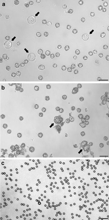
a Three 24-hour interval-old eggplant microspore culture. Cultures at this stage are principally composed of regular eggplant microspores together with few slightly enlarged microspores (arrows). b Eighteen solar day-old eggplant microspore civilisation. Cultures at this stage are principally composed of regular eggplant microspores together with few enlarged microspores or microspore-derived embryos (arrows). c Fluorospheres. These particles are very regular in size and shape. Bars: 50 µm
Precision and accuracy of methods
Equally a preliminary pace to be confident with all the analysis performed, nosotros checked the precision of the pipettes used in this work. The pipetting verification procedure described in Materials and methods yielded %CVs of 0.6 and 0.08% for the 10–100 µl and the 100–grand µl pipette, respectively. These %CVs are notably below the 2% threshold established equally a reference by the manufacturer. Therefore, we causeless that our pipettes were well calibrated, and therefore valid for this study.
We calculated the mean, standard divergence, median and 1st and 3rd quartiles of measurements performed with each method (Table i). Effigy 2a shows a graphical representation of the measurements in cultures at days 3 and eighteen. Next, we performed five independent measurements of fluorosphere suspensions with each method, using the undiluted, 1:2 and 1:10 dilutions. Results are shown in Table ii and represented in Fig. 2b. For both microspore cultures and fluorosphere suspensions, counts with the Neubauer chamber were in general higher than with all other methods, deviating up to ~ fifty% in average from the rest of methods in the instance of fluorospheres. All the means were beneath the theoretical or real density. In terms of precision, the methods based on menstruum cytometry showed the highest values (lowest dispersion) and the Neubauer method showed the highest dispersion of information in microspore measurements (Fig. ii). Nevertheless, the Neubauer method showed a high precision in fluorosphere measurements. The precision of methods that count cells from images taken from the dish (automated and manual-counting methods) presented the lowest values. In terms of accurateness, the Neubauer chamber presented the highest means, therefore closest to the expected values (~ xiii% below in average). Results were remarkably closer to the causeless reference value when undiluted suspensions were measured with the Neubauer method, deviating only 22.5%. Nevertheless, all other methods were markedly beneath, with an average difference of ~ 42%, indicating a low accuracy. In undiluted suspensions, some methods seemed to deviate from the expected value more than at higher dilutions, suggesting that their accuracy may depend on particle density (Fig. 2b). That was the case of paradigm-dependent methods (transmission-counting and automatic counter methods).
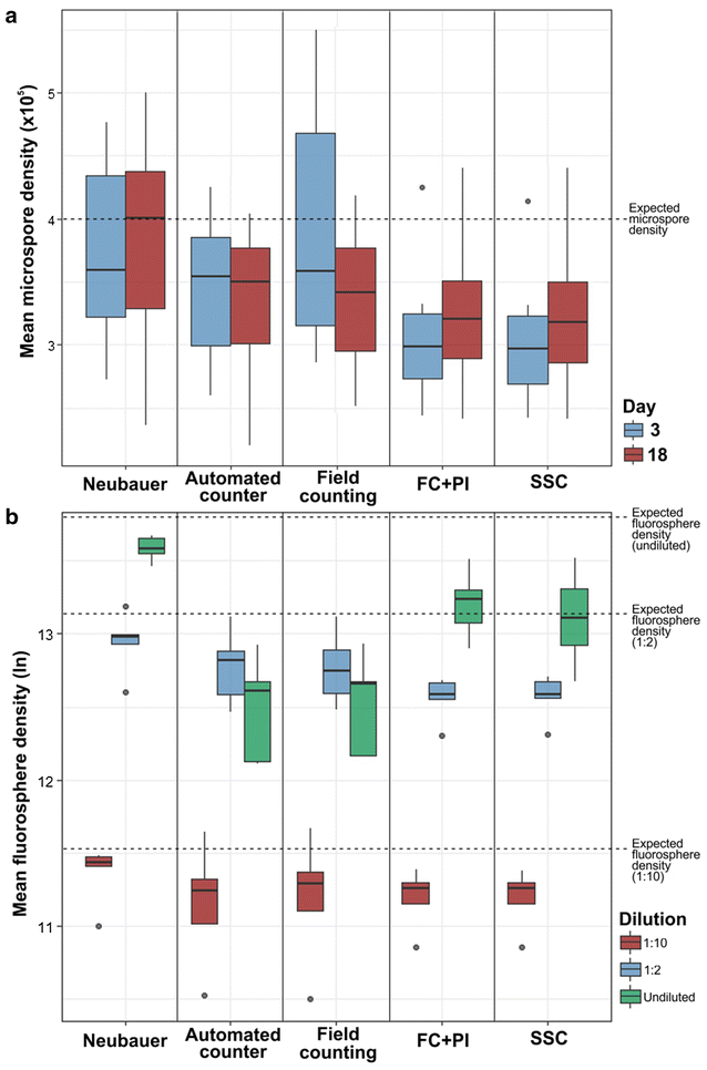
Box-and-whiskers plots for a mean densities of 17 different eggplant microspore cultures measured at days 3 and 18, b fluorospheres at 3 different concentrations: i:10, i:2 and 1:i (undiluted, 1,030,000 microspores/ml). Dashed lines represent the expected microspore (in a) and fluorosphere densities (in b). Annotation that values in B are expressed as neperian logarithms of mean fluorosphere densities. See text for further details
The use of flow cytometry to measure autofluorescence of unstained microspores was the method that showed the worst performance for all parameters tested (Table i). In an attempt to find out the cause of such a discrepancy, we speculated that it could be due to sedimentation of microspores at the bottom of the vial, where the aspiration system cannot attain. To examination this, we designed a set of experiments (see "Methods" section) consisting of recovering the potentially sedimented microspores through successive washing rounds. However, the results of these experiments were non unlike from those without washings (information not shown). Therefore, we concluded that microspore autofluorescence was not sufficiently loftier or homogeneous to detect all the microspores passed through the flow cytometer. As a issue, we decided to discard menstruation cytometry with unstained microspores for further experiments.
Reproducibility of methods
For the analysis of the reproducibility of methods, we compared measurements in samples at two dissimilar moments of culture progression (days 3 and 18). Differences betwixt measurements for each method are depicted by Banal–Altman plots in Fig. iii. As a formal measure, the coefficient of repeatability (CR) was obtained for each method (Table 3) using data of these two different conditions. Moreover, we performed an ANOVA analysis comparing information from both days 3 and xviii where none of the methods showed significant differences (Table 3). Our results showed that both FC + PI (Fig. 3a) and SSC methods (Fig. 3b) were the nigh reproducible, showing narrow limits of understanding and the lowest CR values (Tabular array three). The Neubauer method (Fig. 3c) and the automatic counter (Fig. 3d) appeared equally moderately reproducible, as they presented wider limits of understanding and college CR values (Table 3). Finally, field counting (Fig. 3e) was the least reproducible method. It showed a high CR, the widest limits of understanding, and a bias (~ 35 above) much higher than other methods, where bias ranged betwixt − 17 and 3.
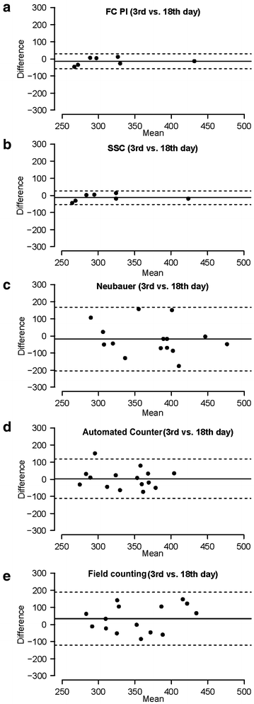
Bland–Altman comparisons of reproducibility of each method by comparing iii and 18 day-old microspore culture data. Difference values of the Y axis are expressed in thousands
Concordance between methods
In order to analyze the level of understanding between methods, we performed Bland–Altman plots from microspore density measurements (Fig. 4). The lowest limits of understanding appeared when Neubauer, SSC and FC + PI methods were compared between them (Fig. 4a–c). SSC and FC + PI (Fig. 4c) showed the highest level of understanding, as revealed past the minimal limits of agreement and non meaning bias of their comparisons. Yet, comparisons between Neubauer and cytometry-based methods (Fig. 4a, b) presented a very loftier bias. Thus, despite their adept level of agreement, flow cytometry methods seem to induce a non-negligible underestimation, at to the lowest degree, with respect to Neubauer method. In general, automated counter and field counting methods showed low concordance with the residuum of methods (Fig. 4d–j), presenting the highest bias when compared to the Neubauer method (Fig. 4d, e), and broad limits of agreement with all methods except between themselves (Fig. 4f). Fluorosphere measurements were not used for this comparison because they accept a known, existent value to compare with.
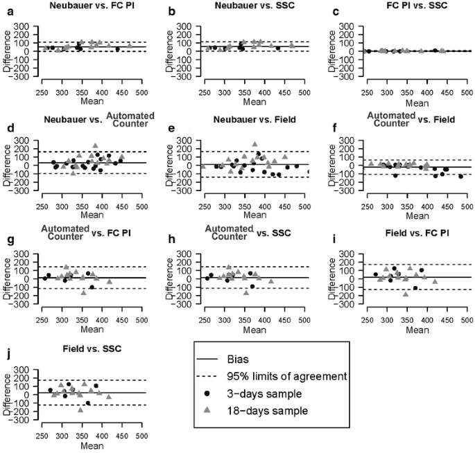
Bland–Altman comparisons of agreement between methods. Difference values of the Y axis are expressed in thousands
Additionally, we assessed agreement betwixt all methods using Lin's concordance correlation coefficient (Tabular array iv). For comparisons between methods, the Lin'south concordance correlation coefficient is preferred over the standard Pearson correlation coefficient considering a Pearson correlation does not detect constant biases, yielding perfect correlations between remarkably differing methods that accept a constant bias. On the other manus, with the Lin's concordance correlation coefficient it is possible to find the presence of constant bias. Using it, we obtained results like to those of the Bland–Altman plots. In general, microspore counts showed in all cases values higher than fluorosphere counts. Concordances between methods were minimal when high fluorosphere concentrations were used, with concordance coefficient values most to zero. The best levels of agreement were found in comparisons between flow cytometry-based methods (cyclopedia coefficient ranging from 0.seven to 1.0) and between field and automatic counter (from 0.v to i). The Neubauer method showed moderate concordance values when compared to flow cytometry methods (from 0.v to 0.viii) except for high fluorosphere concentrations. The lowest level of concordance in microspore counts was found when the field counting method was compared with the Neubauer, PI and SSC.
Aligning of the initial density with different counting methods
The results presented above were based on microspore cultures whose density was initially adapted to 400,000 microspores/ml using the Neubauer method. All these results revealed a positive bias of the Neubauer method with respect to nigh of the other methods (Fig. 4). In order to double cheque this surprising observation, we performed 12 new cultures and adapted their initial densities using the Neubauer, cell counter, field and FC + PI counting methods (3 cultures adjusted with each method). Since previous results of FC + PI and SSC showed a most verbal match (Fig. 4j), nosotros omitted the use of SSC for these experiments, bold practical equivalence of both methods. Then, we checked culture densities at 3 and 18-day sometime cultures using each of the four methods, every bit usual. Equally seen in Fig. 5, an initial aligning to 400,000 microspores/ml with the Neubauer method fabricated that three and 18-day measurements with the aforementioned method were notably similar (two.7 and 4.8% deviation), but ix.2–21.1% higher than those of the other methods. When the automated jail cell counter was used for the initial adjustment, all counts (with the exception of the automated counter) were above 400,000, being notably higher in the example of the Neubauer method (26.8 and 29.one% for 3 and xviii-mean solar day one-time cultures, respectively). An initial adjustment with the field counting method resulted in values around 400,000 when counted with the Neubauer method, but 8.6–21.1% lower when counted with the other three methods, including the initial (field counting). When cultures were initially adjusted with the menstruation cytometer, counts at days iii with all four methods revolved around 400,000, but they were clearly below at days 18, except for the Neubauer method. Yet, a trend to underestimate its own initial count was found for flow cytometry at both timepoints. From these results, we could conclude that the automated counter, field counting and menstruum cytometry tended to underestimate densities, since when they were used to arrange the initial density, all other methods yielded higher counts at both timepoints. This was particularly dramatic in the case of menstruum cytometry and field counting, which at days three and xviii yielded counts lower than in the initial adjustment made using the same methods. As to the Neubauer method, it could be thought that it tends to overestimate densities, since in general, this was the method yielding highest counts. However, it must be noted that in most cases, the Neubauer method showed the counts closest to the expected value of 400,000 microspores/ml.
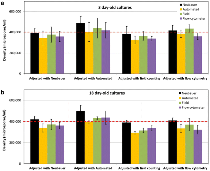
Comparing of microspore density measurements performed at days 3 (a), 18 (b) with iv counting methods in cultures whose initial microspore density was adjusted to 400.000 microspores/ml (cherry-red dashed line) with each method. Run into text for further details
Microspore density estimation from fluorosphere counting in mixed samples
The common use of fluorospheres to estimate cell densities implies their use as internal standards mixed with cell suspensions, unremarkably for flow cytometry. In our study, we also tried to gauge microspore density from a known quantity of fluorospheres mixed with cells. Measurements were performed with Neubauer, automated counter, field counting and SSC method. FC + PI method was omitted because of its similarity with SSC. Results of these assays are shown in Table 5. The automated counter paradigm detection arrangement was unable to differentiate between fluorospheres (smaller) and microspores (larger), so we could not obtain any estimation in this case. For the remainder of cases, estimations of microspore density were always higher up the theoretical value, ranging from 19 to 37% of difference. Thus, we concluded that at least for microspores, this method, although fast and straightforward, is not accurate enough, at least when used with non flow cytometry methods. Indeed, the all-time results were obtained with the latter, which is reasonable since this counting strategy has been designed for flow cytometry.
Discussion
For most jail cell culture systems, optimal cell progression depends on the optimization of the initial cell density at the onset of the civilization. A paradigmatic example of this is isolated microspore civilisation, where the developmental switch relies on the successful optimization of many dissimilar experimental parameters that critically affect the efficiency of the process, and one of them is the density at which microspores are suspended in liquid medium. It affects non simply the efficiency of the induction of microspores towards embryogenesis, just also a successful conversion of induced microspores into viable, germinating embryos [three, v, 10, sixteen, 37, 38]. Due to the importance of this initial step, non only for microspore culture but for virtually all animal and plant cell cultures, in this work nosotros compared the use of different methods that have been used or could potentially be used to calculate particle densities using ii different particles: eggplant microspores and fluorospheres. In lite of our results, we can dissever the methods used in 3 groups: flow cytometry methods, automated counter and field counting, and Neubauer bedroom. Each has both positive and negative aspects. They are summarized in Table vi and discussed below.
Menstruum cytometry methods are the most reproducible, simply they have depression accuracy and precision
We evaluated three different methods based on the apply of menstruum cytometry. The commencement method consisted on the assay of unstained microspores, assuming that the natural autofluorescence of the exine glaze could be sufficiently high to exist detected and quantified by the system. Nonetheless, the analysis of vii three-24-hour interval old cultures was enough to realize that this method presented serious limitations. It seemed that exine autofluorescence is not sufficiently intense and/or homogeneous to be detected in all the microspores, at least in our eggplant microspore cultures. Obviously, nosotros strongly discourage its use.
The FC + PI method, together with SSC, provided the almost reproducible results, despite the different concrete principles used to identify and count flowing microspores. FC + PI and SSC exhibited almost identical results in all the experiments and statistical tests performed. Nevertheless, evaluation of accuracy (using a standard with a known concentration) and precision (with a high number of measurements) pb u.s. to conclude that cytometry methods are non as accurate and precise as the rest. In add-on, they repeatedly showed a negative bias with respect to other methods, specially the Neubauer method. Thus, a question arises as to why the flow cytometry methods nosotros used have such a low performance. The use of fluorescent chaplet is a well-known method to calculate cell densities through period cytometry, at to the lowest degree for animal cell cultures [39, twoscore]. Our work is non the only one showing a consistent bias (either positive or negative) of the Neubauer method compared to others [41]. Yet, other studies have compared hemacytometers versus automated counting methods in animal cells [12, 39, 40, 42,43,44,45], and no significant bias has been reported. After a thorough study of the different user-based technical factors that could potentially cause such a bias (including bad calculations, incorrect chamber dimensions, pipetting errors, uneven cell distribution, contamination, user-to-user variation, and filling issues, among others), nosotros found a possible crusade that might explain such a bias. Several studies comparing methods to summate the flow rate of flow cytometer fluidics systems, have acknowledged the limitations of many catamenia cytometers to perform a proper interpretation of the volume loaded [46, 47], which may preclude a correct interpretation of particle densities. This led u.s.a. to evaluate the volume interpretation accurateness of our menses cytometer and, as expected, the effective volume loaded never coincided with the volume estimated past the device (data at present shown). This could surely explain the negative bias and the low accurateness and precision of the flow cytometry-based methods we used.
On the other hand, flow cytometers are expensive systems, even in their bones, compact versions. As to FC + PI, a CyFlow cytometer equipped with a Nd-YAG green laser for PI is around €35,000. It is possible to apply other fluorescent stains excitable past UV LED calorie-free sources, which are cheaper than Nd-YAG dark-green lasers. For example, the CellTracker Blue CMAC Dye from Molecular Probes. Still, although UV LED light sources could drop the price of this system down to ~ €29,000, this alternative may all the same exist expensive for many research groups. Another limitation of this method is the need for staining. We used 1 h to ensure a complete and reproducible PI staining. Perhaps, this time could be optimized trying different combinations of incubation fourth dimension and PI concentration, or even other dyes. Anyway, a staining step volition always exist needed, which may slow down the whole process considerably.
The third catamenia cytometry-based method tested was SSC analysis. Although we performed this analysis with PI-stained samples, the physics of this method allows for an interpretation of the internal complexity of individual particles independent of their fluorescent emission. This means that the time-consuming staining step could be avoided, needing only few min to analyze tens of thousands cells. Withal, it must be noted that the basic equipment needed to perform this blazon of analysis has an estimated circular cost revolving around €35,000. A like model with two light sources (a Nd-YAG light-green laser + a UV LED lamp) may exist around €40,000. Clearly, this equipment may not be routinely available for all cell civilization laboratories. Alternatively, the user might carry civilisation vessels to a flow cytometry facility. Notwithstanding, this would imply long times and potential risks for cultured cells, perhaps incompatible with routine prison cell culturing. Clearly, we discourage this method for that purpose.
Automated counter and field counting methods accept problems associated with image acquisition
The second group of methods tested was field and automatic counting. The working principle of both methods is like, but as a departure, the field counting method needs merely a microscope, and is largely based on an interaction with the user, who must acquire the images or observe microscopic fields, and then count all the particles observed. In principle, information technology could be thought that a human eye would be more authentic than a machine for cell identification and counting. The different comparisons of the automated counter with field counting revealed that in full general, results are very similar between them, which indicates that automated image analysis is at least every bit correct every bit man observations. In addition, user-based methods use to be time-consuming and subjected to user bias and putative lack of expertise. Therefore, nosotros postulate that, despite their reduced price and ease of use, the methods that imply a higher user interaction should be avoided in order to increase accuracy and reduce the experimental variation between cultures.
On the other paw, these two methods showed a low accuracy and a moderate precision (see Tabular array 6). In our experience, we accept detected some image acquisition issues that might explain it. First of all, nosotros have consistently observed that cells and fluorospheres do not distribute homogeneously in the civilisation dish, which may brand mandatory a software-based tool to recoup for such uneven distribution. The second reason could exist automatic focusing, which is common to most image-based automatic counting devices. The algorithm of the equipment nosotros used searches the most contrasted area in the z-centrality and takes pictures of it. The problem appears when not all particles are well focused or they are in unlike focal planes, which precludes their proper identification. Moreover, in the case of fluorospheres, suspended in a viscous solution, long times are needed for them to settle downwardly. In some cases, they practice non fifty-fifty settle downwardly, and stay floating. Summarizing, automated and manual counting methods may not exist the best selection to approximate prison cell or particle suspension densities because they are prone to miss particles out of the focal aeroplane.
The estimated price of the automated counter we used is effectually €15,000–€xx,000. Still, it must be noted that it includes a born microscope. Other, basic versions of this system need a microscope to be coupled to, merely they are much cheaper, which makes it more convenient when the laboratory is already equipped with a light microscope. In one case installed and calibrated, it is a rather straightforward and easy-to-use arrangement that allows for quick measurements of cell densities. Other systems such every bit the Cellometer Auto T4 (Nexcelom Biosciences) have a congenital-in CCD chip in society to load, paradigm and analyze the sample in the aforementioned machine, or use non-epitome-based methods to count cells, such equally the Scepter Cell Counter from Millipore, which uses the impedance-based Coulter principle to observe cells. This avoids the need for a microscope, but for some microscope-independent systems, the cost is similar to that of a microscope + a microscope-coupled automatic counter. In improver, Coulter-based systems are applicative only to a limited range of particle sizes. All this considered, automated cell counters announced as a more than affordable alternative to flow cytometers.
The Neubauer sleeping accommodation showed the best overall performance
As mentioned in the introduction, the methods based on the apply of counting chambers appear as the nigh popular and widely used to calculate jail cell densities. Most probable, this is due to its affordability. Indeed, this method requires simply a Neubauer sleeping room (around €260) and a basic microscope, bachelor in well-nigh laboratories. There may also be a general assumption that, since the employ of these methods is widely extended, they must be sufficiently well known and therefore, accurate and reliable. After many different experiments, using both microspores and fluorospheres, the Neubauer method repeatedly showed a positive bias with respect to the other methods used, merely its means were always virtually the theoretical value, whereas the other methods were e'er below. In addition, analysis of accuracy and precision demonstrated that it is the nearly reliable method on the basis of its depression dispersion and high accurateness. However, information technology must exist noted that the number of cells counted in this work is much higher than that of common routine counts, which surely compensated for the very dissimilar results we observed in individual data from each bedroom grid (data non shown). Due to this, a major limitation of this method might exist the reduced number of cells counted in routine procedures. However, it can exist hands overcome by increasing the amount of counted particles.
Conclusions
Based on our results, it seems evident that amongst the methods used, those based on menses cytometry are, past far, the most reproducible and concordant, but the worst in terms of accuracy and precision, likely due to an improvable period rate measurement. Automatic counter and field counting methods showed a low accurateness but a moderate precision. Their actual problem relates to the prototype conquering system, improvable too. Perhaps the most important determination of this work is that counting chambers and in particular Improved Neubauer chambers are the most reasonable option to routinely mensurate jail cell densities. This is of import since their use is widely extended among the research community, but at that place are not abundant comprehensive comparisons to support its apply from a technical perspective. In most cases, the reason for this adoption has been "because it was used previously in the protocol nosotros are applying". Yet, our advice to hereafter users of Neubauer chambers would exist to increment the number of cells counted in each assay, in order to reach these standards of accuracy and low dispersion.
References
-
Kobayashi T, Higashi Yard, Saitou T, Kamada H. Physiological properties of inhibitory conditioning factor(south), inhibitory to somatic embryogenesis, in high-density cell cultures of carrot. Plant Sci. 1999;144:69–75.
-
Schween K, Hohea A, Koprivova A, Reski R. Effects of nutrients, jail cell density and civilization techniques on protoplast regeneration and early protonema development in a moss, Physcomitrella patens. J Plant Physiol. 2003;160:209–12.
-
Kim M, Jang I-C, Kim J-A, Park East-J, Yoon Grand, Lee Y. Embryogenesis and institute regeneration of hot pepper (Capsicum annuum L.) through isolated microspore culture. Plant Jail cell Rep. 2008;27:425–34.
-
Hoekstra S, Vanzijderveld MH, Heidekamp F, Vandermark F. Microspore culture of Hordeum vulgare L.—the influence of density and osmolality. Plant Cell Rep. 1993;12:661–5.
-
Castillo AM, Valles MP, Cistue L. Comparison of anther and isolated microspore cultures in barley. Furnishings of culture density and regeneration medium. Euphytica. 2000;113:i–8.
-
Huang B, Bird S, Kemble R, Simmonds D, Keller W, Miki B. Effects of civilisation density, conditioned medium and feeder cultures on microspore embryogenesis in Brassica napus L. cv. Topas. Constitute Cell Rep. 1990;eight:594–7.
-
Li HC, Devaux P. High frequency regeneration of barley doubled haploid plants from isolated microspore culture. Found Sci. 2003;164:379–86.
-
Kott LS, Polsoni L, Ellis B, Beversdorf WD. Autotoxicity in isolated microspore cultures of Brassica napus. Tin can J Bot. 1988;66:1665–70.
-
Raina SK, Irfan ST. High-frequency embryogenesis and plantlet regeneration from isolated microspores of indica rice. Plant Cell Rep. 1998;17:957–62.
-
Ma R, Guo YD, Pulli S. Comparison of anther and microspore civilization in the embryogenesis and regeneration of rye (Secale cereale). Constitute Prison cell Tissue Organ Cult. 2004;76:147–57.
-
Höfer M. In vitro androgenesis in apple—improvement of the induction stage. Establish Prison cell Rep. 2004;22:365–70.
-
Ongena K, Das C, Smith JL, Gil Southward, Johnston Grand. Determining cell number during prison cell culture using the scepter cell counter. J Vis Exp. 2010;45:2204.
-
Corral-Martínez P, Parra-Vega V, Seguí-Simarro JM. Novel features of Brassica napus embryogenic microspores revealed past high pressure freezing and freeze commutation: testify for massive autophagy and excretion-based cytoplasmic cleaning. J Exp Bot. 2013;64:3061–75.
-
Regner F. Anther and microspore culture in Capsicum. In: Jain SM, Sopory SK, Veilleux RE, editors. In vitro haploid production in higher plants, vol. iii. Dordrecht: Kluwer; 1996. p. 77–89.
-
Nelson Thousand, Mason A, Castello M-C, Thomson L, Yan G, Cowling W. Microspore culture preferentially selects unreduced (2n) gametes from an interspecific hybrid of Brassica napus Fifty. ×Brassica carinata Braun. Theor Appl Genet. 2009;119:497–505.
-
Kim 1000, Park Due east-J, An D, Lee Y. High-quality embryo production and plant regeneration using a 2-step culture system in isolated microspore cultures of hot pepper (Capsicum annuum L.). Plant Cell Tissue Organ Cult. 2013;112:191–201.
-
Simmonds DH, Long NE, Keller WA. Loftier plating efficiency and establish regeneration frequency in low density protoplast cultures derived from an embryogenic Brassica napus prison cell suspension. Plant Cell Tissue Organ Cult. 1991;27:231–41.
-
Gémes Juhász A, Kristóf Z, Vági P, Lantos C, Pauk J. In vitro anther and isolated microspore culture as tools in sweet and spice pepper breeding. Acta Hort. 2009;829:61–4.
-
Lantos C, Juhasz AG, Vagi P, Mihaly R, Kristof Z, Pauk J. Androgenesis induction in microspore civilization of sweet pepper (Capsicum annuum L.). Found Biotechnol Rep. 2012;half dozen:123–32.
-
Nageli M, Schmid JE, Postage stamp P, Buter B. Improved formation of regenerable callus in isolated microspore culture of maize: touch on of carbohydrates, plating density and time of transfer. Plant Jail cell Rep. 1999;xix:177–84.
-
Gu HH, Zhou WJ, Hagberg P. High frequency spontaneous production of doubled haploid plants in microspore cultures of Brassica rapa ssp chinensis. Euphytica. 2003;134:239–45.
-
Rudolf K, Bohanec B, Hansen M. Microspore civilisation of white cabbage, Brassica oleracea var. capitata L.: Genetic improvement of not-responsive cultivars and result of genome doubling agents. Constitute Breed. 1999;118:237–41.
-
Sato S, Katoh N, Iwai Southward, Hagimori M. Frequency of spontaneous polyploidization of embryos regenerated from cultured anthers or microspores of Brassica rapa var. pekinensis L. and B. oleracea var. capitata L. Breed Sci. 2005;55:99–102.
-
Nicoloso FT, Val J, Vanderkeur Grand, Vaniren F, Kijne JW. Flow-cytometric jail cell counting and Dna estimation for the study of plant cell population dynamics. Plant Cell Tissue Organ Cult. 1994;39:251–9.
-
Schulze D, Pauls KP. Flow cytometric characterization of embryogenic and gametophytic development in Brassica napus microspore cultures. Institute Cell Physiol. 1998;39:226–34.
-
Schulze D, Pauls KP. Flow cytometric analysis of cellulose tracks development of embryogenic Brassica cells in microspore cultures. New Phytol. 2002;154:249–54.
-
Corral-Martínez P, Seguí-Simarro JM. Efficient product of callus-derived doubled haploids through isolated microspore civilisation in eggplant (Solanum melongena L.). Euphytica. 2012;187:47–61.
-
Corral-Martínez P, Seguí-Simarro JM. Refining the method for eggplant microspore civilisation: effect of abscisic acrid, epibrassinolide, polyethylene glycol, naphthaleneacetic acid, 6-benzylaminopurine and arabinogalactan proteins. Euphytica. 2014;195:369–82.
-
Rivas-Sendra A, Corral-Martínez P, Camacho-Fernández C, Seguí-Simarro JM. Improved regeneration of eggplant doubled haploids from microspore-derived calli through organogenesis. Plant Cell Tissue Organ Cult. 2015;122:759–65.
-
Rivas-Sendra A, Campos-Vega M, Calabuig-Serna A, Seguí-Simarro JM. Development and characterization of an eggplant (Solanum melongena) doubled haploid population and a doubled haploid line with loftier androgenic response. Euphytica. 2017;213:89.
-
Salas P, Rivas-Sendra A, Prohens J, Seguí-Simarro JM. Influence of the stage for anther excision and heterostyly in embryogenesis consecration from eggplant anther cultures. Euphytica. 2012;184:235–50.
-
Hervás D: random_path: Shortest route for random sampling in circles. ZENODO; 2014.
-
Shapiro H. Practical menses cytometry. 3rd ed. New York: Alan R. Liss; 1994.
-
Banal JM, Altman DG. Measuring agreement in method comparison studies. Stat Methods Med Res. 1999;8:135–sixty.
-
Lin LI. A concordance correlation coefficient to evaluate reproducibility. Biometrics. 1989;45:255–68.
-
R Development Core Squad. A language and environment for statistical computing. Vienna: The R Foundation for Statistical Calculating; 2011.
-
Reynolds TL. Pollen embryogenesis. Constitute Mol Biol. 1997;33:1–x.
-
Wang M, van Bergen S, Van Duijn B. Insights into a primal developmental switch and its importance for efficient institute breeding. Plant Physiol. 2000;124:523–30.
-
Cadena-Herrera D, Esparza-De Lara JE, Ramírez-Ibañez ND, López-Morales CA, Pérez NO, Flores-Ortiz LF, Medina-Rivero E. Validation of three feasible-prison cell counting methods: manual, semi-automated, and automated. Biotechnol Rep. 2015;vii:nine–sixteen.
-
Huang L-C, Lin W, Yagami M, Tseng D, Miyashita-Lin E, Singh N, Lin A, Shih S-J. Validation of cell density and viability assays using Cedex automatic cell counter. Biologicals. 2010;38:393–400.
-
Bailey Due east, Fenning N, Chamberlain S, Devlin L, Hopkisson J, Tomlinson M. Validation of sperm counting methods using limits of agreement. J Androl. 2007;28:364–73.
-
Johnston One thousand. Automated handheld instrument improves counting precision across multiple cell lines. Biotechniques. 2010;48:325–7.
-
Louis KS, Siegel AC. Prison cell viability analysis using trypan blue: manual and automated methods. In: Stoddart MJ, editor. Mammalian prison cell viability, vol. 740. Humana Press: New York; 2011. p. 7–12 (Methods in molecular biological science).
-
Tucker KG, Chalder S, Al-Rubeai Thou, Thomas CR. Measurement of hybridoma cell number, viability, and morphology using fully automated image assay. Enzyme Microb Technol. 1994;16:29–35.
-
Collins CE, Immature NA, Flaherty DK, Airey DC, Kaas JH. A rapid and reliable method of counting neurons and other cells in brain tissue: a comparing of menstruum cytometry and manual counting methods. Front end Neuroanat. 2010;4:5.
-
Marie D, Simon N, Vaulot D. Phytoplankton Cell Counting Past Menses Cytometry. In: Alndersen RA, editor. Algal culturing techniques, vol. 17. Burlington: Elsevier; 2005. p. 253–67.
-
Storie I, Sawle A, Goodfellow G, Whitby 50, Granger Five, Ward RY, Skin J, Smart T, Reilly JT, Barnett D. Perfect count: a novel arroyo for the single platform enumeration of absolute CD4 + T-lymphocytes. Cytometry Function B: Clinical Cytometry. 2004;57B:47–52.
Authors' contributions
CCF generated the experimental piece of work, analyzed the results, and contributed to manuscript confection. DH developed all statistical analyses. ARS contributed to the generation of experimental work and manuscript confection. MPM contributed to the experimental blueprint and results discussion; JMSS designed the experimental piece of work, analyzed the results, and wrote the manuscript. All authors read and approved the final manuscript.
Acknowledgements
Not applicative.
Competing interests
The authors declare that they take no competing interests.
Availability of data and materials
Data sharing is not applicable to this article as no datasets were generated or analysed during the electric current study.
Consent for publication
Not applicable.
Ethics approval and consent to participate
Not applicative.
Funding
This work was supported by Grant UPV-Iron-2013-7 from Universitat Politècnica de València and Hospital Universitari i Politècnic La Fe to JMSS and MPM, and Grants AGL2014-55177-R and AGL2017-88135-R to JMSS from Spanish Ministerio de Economía y Competitividad (MINECO) jointly funded by FEDER. CCM and ARS are recipients of PhD Fellowships from Generalitat Valenciana and Universitat Politècnica de València, respectively.
Publisher'south Notation
Springer Nature remains neutral with regard to jurisdictional claims in published maps and institutional affiliations.
Author information
Authors and Affiliations
Corresponding author
Additional files
Rights and permissions
Open Access This article is distributed under the terms of the Creative Commons Attribution four.0 International License (http://creativecommons.org/licenses/by/4.0/), which permits unrestricted use, distribution, and reproduction in any medium, provided you give appropriate credit to the original author(s) and the source, provide a link to the Artistic Eatables license, and indicate if changes were made. The Creative Commons Public Domain Dedication waiver (http://creativecommons.org/publicdomain/zero/1.0/) applies to the information made available in this article, unless otherwise stated.
Reprints and Permissions
About this article
Cite this article
Camacho-Fernández, C., Hervás, D., Rivas-Sendra, A. et al. Comparison of six different methods to calculate cell densities. Establish Methods xiv, 30 (2018). https://doi.org/ten.1186/s13007-018-0297-4
-
Received:
-
Accepted:
-
Published:
-
DOI : https://doi.org/10.1186/s13007-018-0297-four
Keywords
- Automated cell counter
- Cell counting
- Flow cytometry
- Fluorospheres
- Hemacytometer
- Image analysis
- Microscopy
- Microspore culture
Source: https://plantmethods.biomedcentral.com/articles/10.1186/s13007-018-0297-4
Posted by: hagemanhimpre.blogspot.com

0 Response to "How To Calculate The Number Of Cell An Animal Has"
Post a Comment